Protein extraction
Protein extraction is a key step for analytical methods such as Western Blot and enzymatic assays. Below, Bertin’s top scientists have shared best practices to improve your homogenization protocol for a wide range of tissues, including tips to preserve the integrity of the proteins in your samples and achieve optimal yield.
Proteins are macromolecules consisting of long chains of amino acids. They include enzymes, cell signaling proteins, ligand binding proteins, and cytoskeletal proteins. Protein extraction is the first step for most proteomics research methods (e.g. Western Blot, SDS-PAGE, mass spectrometry) as well as protein purification. Many techniques have been developed to maximize yield and purity for samples such as tissues or cultured cells. To get a high protein yield, samples (e.g. plant and animal tissues, cultured cells) must be homogenized uniformly and efficiently. This can be challenging as many samples contain protease protein-degrading enzymes as well as other interfering substances (e.g. keratins, phenolics, lipids, and organic acids). Thanks to 3D bead-beating technology — the gold standard for mechanical disruption — the Precellys homogenizers can homogenize virtually any type of tissues or cells and ensure that researchers always get the highest protein yield and purity.
Below, our top scientists have shared best practices for homogenization with the Precellys products range to maximize the recovery of high-quality proteins.
Tips for a successful protein extraction
Choice of buffer and ratio mg tissue/volume of lysis buffer
The composition of the homogenization buffer should be adjusted according to the sample type, the location of the protein of interest, the required yield, and the nature of the downstream analysis. In general, a RIPA buffer can be a good starting point for optimization (150 mM NaCl, 1% NP-40, 0.25% Na-deoxycholate, 50 mM Tris-HCl (pH 7.4)). Depending on the assays, detergents such as SDS can be added to the buffer to improve the recovery of soluble protein.
Depending on downstream application the addition phosphatase and proteases inhibitors is advised to prevent protease-mediated protein degradation and/or dephosphorylation. Bertin’s Life Sciences team has developed the Protein safe kits, a state-of-the-art lysing kit containing a proprietary mix of protease inhibitors (see Figure 2). It should be noted that for some applications such as mass spectrometry, protease inhibitors should be used with great caution as they can interfere with results. We recommend using our standard lysing kits rather than the Protein Safe kits in the case of mass spectrometry workflows.
Another important factor is finding a good ratio (mg tissue/µl lysis buffer) between the quantity of tissue to homogenize and the volume of lysis buffer added in the homogenization tube. Overloading tissue in the tube could lead to poor or incomplete homogenization. On the other hand, adding too much lysis buffer will result in over-diluted non-exploitable samples. Depending on the tissue sample you are working on, a good starting point for the tissue/buffer ratio can be found in the table below.
| Tissue sample | Weight mg | Volume μl | Lysing kit | Tube format | Hints |
|---|---|---|---|---|---|
| Soft tissue: Liver, Brain, Spleen | 80-100 mg | 700 μl – 1ml | CK14 | 2 ml | Short homogenization cycles |
| Elastic tissue (skin, intestine) and tumors | 30-50 mg | 500 μl | CK28R, CKMix50R R = reinforced tubes |
2 ml | *** Do not submit elastic tissue and tumors as whole chunk, chop of tissue with scalpel in small pieces for good quality homogenates** |
| Hard tissue. Bones, cartilage | 50-100 mg | 700 μl | CK28R, CKMix50, MK28R | 2 ml |
Temperature control
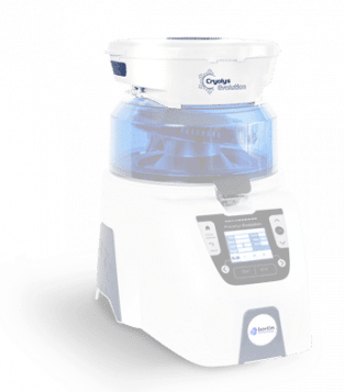
Proteins are thermosensitive molecules, and as such, should be protected against excessive heating during homogenization. Consequently, protocols combining adapted homogenization speeds with short homogenization time (<15s) and lower cycles should be preferred. The Cryolys Evolution allows a precise control of samples temperature during each Precellys run.
Tips for temperature control
Before the homogenization, we recommend placing your Precellys tubes on ice and chilling your lysis buffer. For protein work, it is preferable to have breaks of at least one minute between homogenization cycles, or to use the Cryolys cooling unit at +4°C.
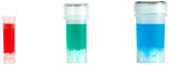
When setting up the homogenization of your samples for the first time, it is good to visually checking the homogenization efficiency after the second cycle by looking at the consistency of the homogenate in the tube. Once the liquid inside the tube is homogeneous, the homogenization cycles should be stopped.
Important: Do not freeze Precellys tubes with dry ice or liquid nitrogen prior to homogenization as it fragilizes the tube material and could result in them breaking during the homogenization process.
Discover the protein safe kit in video
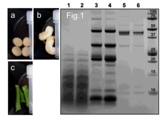
Figure 1: SDS-PAGE analysis of food extracts homogenized with Precellys Evolution, with or without PBS pH7.4 addition.
1 – dog biscuits homogenized with PBS pH7.4;
2 – dog biscuits homogenization. without PBS pH7.4;
3 – cashew nuts homogenization with PBS pH7.4;
4 – cashew nuts homogenization without PBS pH7.4;
5 – green beans homogenization with PBS pH7.4;
6 – green beans homogenization without PBS pH7.4.
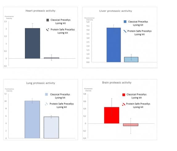
Figure 2: Impact of the Protein Safe Lysing kit on protease activity during animal tissues homogenization with Precellys.
The different tissue types (liver, brain, heart and lung) were cut off in pieces. 50mg of sample were then loaded in either CK14 Protein Safe lysing tubes or in regular CK14 Lysing tubes. Volume was then completed with 1,6mL of buffer. Tubes were placed on the Precellys® Evolution equipped with Cryolys® Evolution instrument and processed using a generic homogenization program. 400µl of the obtained homogenate were then transferred in a new tube to proceed to proteases activity assay. Assay is based on incubation of the homogenate with a universal proteases resofurin labelled substrate. Active proteases degrade this substrate and release a fluorescent molecule. The level of fluorescence detected on the sample by fluorimeter is then related to protease activity. In case proteases are inactive (Protein Safe inhibitor), the reaction cannot occur and no or low fluorescence is observed. Fluorescence levels reflecting the proteases activity levels were measured in the different samples following the method described previously. The different activity levels measured for a same sample extracted with Classical Precellys® Lysing kit or the Protein Safe Lysing kit are represented in the figures 1 to 4 for Heart, Liver, Lung and Brain.
Related products