When Your Animal Model Changes, Why Change Your Antibody?
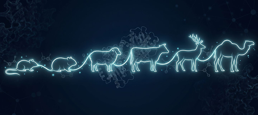
Prion disease research is no longer limited to traditional models such as mice and hamsters. Today, studies and surveillance programmes for transmissible spongiform encephalopathies (TSEs) also involve cattle, small ruminants, cervids and camelids.
This diversification raises an important question: will your antibody still recognise its target when you switch animal models?
The anti-PrP monoclonal antibodies SAF32 (A03202) and Sha31 (A03213) provide a practical answer. In the published studies reviewed, these two clones have been used to detect the PrP protein in 10 species belonging to 5 mammalian orders, ranging from primates to marsupials (1-12).
Two Clones, Broad Species Coverage
Published studies document the use of SAF32 and Sha31 across 10 mammalian species from 5 distinct taxonomic orders, including humans, mice, hamsters, sheep, cattle, white-tailed deer and dromedary camels (1-12).
This breadth of species coverage is an advantage for laboratories working with multiple animal models or conducting comparative studies.
SAF32: Targeting the N-terminal Region of PrP
The monoclonal antibody SAF32 (A03202) recognises the octapeptide repeat region located within the N-terminal portion of the PrP protein. The scientific literature reports its use in several species, including humans (9, 10), mice (8), dromedary camels (1) and the red-tailed phascogale (Phascogale calura), an Australian marsupial (4).
Sha31: A Widely Used Antibody for Cross-Species Detection
The Sha31 (A03213) antibody targets the α1 helix within the C-terminal globular domain of the PrP protein. The epitope sequence recognised by Sha31 (YEDRYYRE) has been described as conserved across multiple species (11), helping to explain its widespread use in PrP detection studies and TSE surveillance programmes.
The literature reports its use in humans, mice, hamsters and sheep (12), cattle (3, 7), white-tailed deer and raccoons (2), bank voles (5) and dromedary camels (1).
An Advantage for Multi-Species Studies
This documented species coverage helps minimise reagent changes when moving from one animal model to another and facilitates comparative studies by enabling the use of the same detection antibody throughout a project. It also reduces a potential source of experimental variability and supports more consistent comparisons between results obtained within the same study.
This is particularly valuable for laboratories involved in prion research, molecular strain typing and TSE surveillance programmes.
Versatility Demonstrated in the Scientific Literature
Several publications highlight the ability of these antibodies to perform across a range of experimental settings:
- Vilette et al. (2016): Sha31 was used to detect PrPres from multiple species within a single study (12).
- Cassmann et al. (2022): Sha31 was employed in studies investigating Chronic Wasting Disease (CWD) transmission in white-tailed deer (2).
- Amara et al. (2025): SAF32 and Sha31 were used together in the first identification of camel prion disease in Tunisia. Sha31 was used for PrPres detection, SAF32 for the molecular characterisation distinguishing this disease from classical scrapie (1, preprint).
Are You Working With a New Species?
Explore the documented species coverage for SAF32 (A03202) and Sha31 (A03213) and choose the anti-PrP antibody best suited to your application.
Download the Species Coverage Guide